Featured Works
Recently Uploaded
Featured Researcher
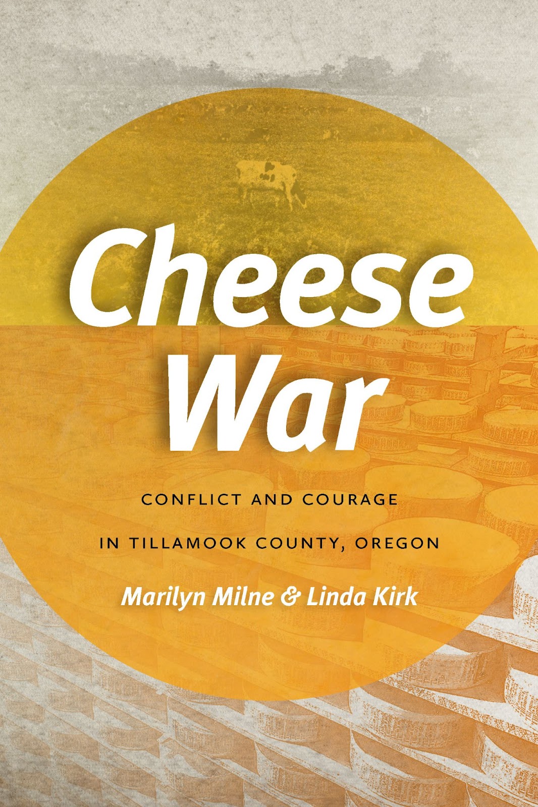
Twenty OSU Press new releases are now available for the OSU community on ScholarsArchive. Titles include Cheese War: Conflict and Courage in Tillamook County, Oregon by sisters Marilyn Milne and Linda Kirk and Dead Wood: The Afterlife of Trees by Ellen Wohl.
You can see other OSU Press titles on ScholarsArchive here.

More information about the OSU Press is available on the Press website.